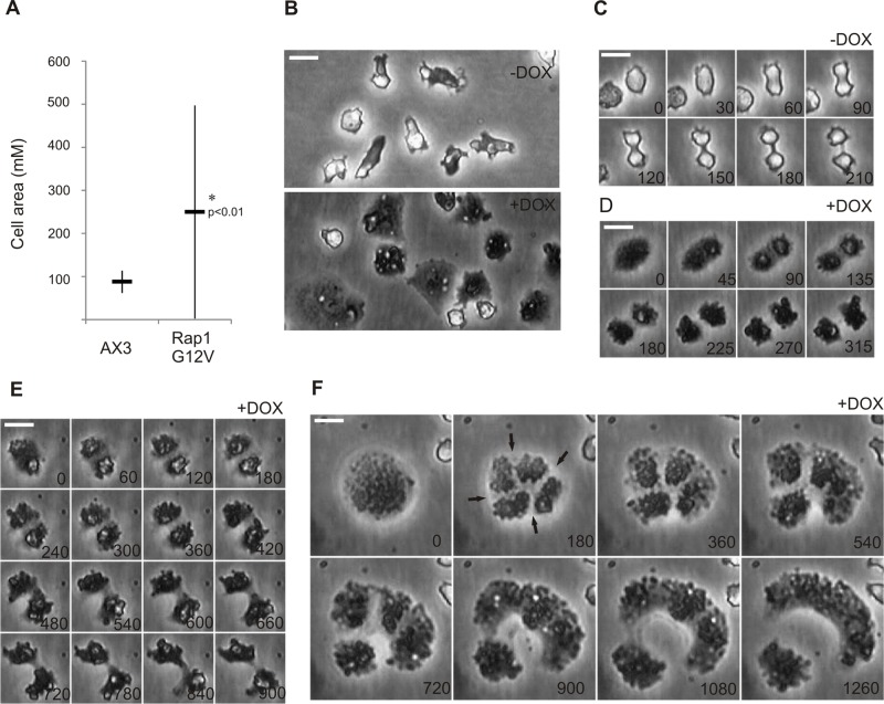
FIGURE 2:
Hyperactivation of Rap1 leads to various cytokinetic defects on substrates. Dictyostelium Rap1G12V (+DOX) and control (−DOX) cells. Shown are (A) graph depicting the average cell size of vegetative grown cells and (B–F) images of cells undergoing cytokinesis on substrates; time in seconds is indicated. Arrows in F show the regions where the furrow is being formed. Scale bars: 10 μm. * indicates significantly different from wild type with p < 0.01.

