FIGURE 4:
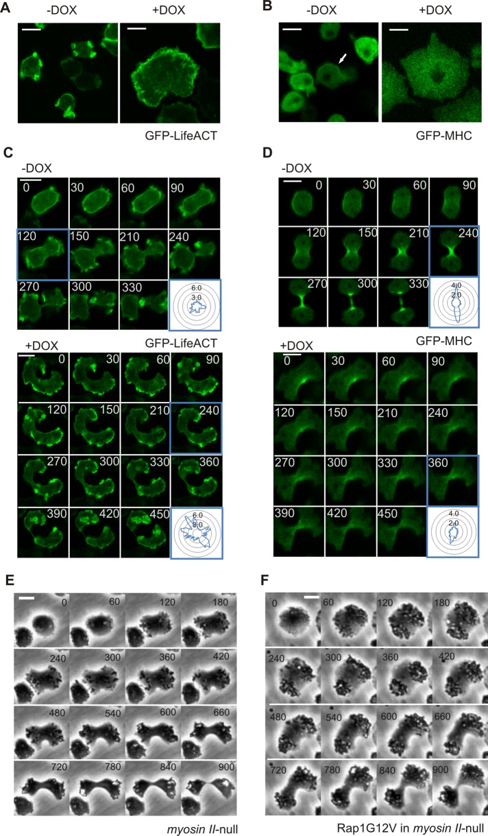
Rap1 regulates myosin and actin cytoskeleton dynamics. Images of vegetative induced (+DOX) or control (−DOX) cells expressing (A) Lifeact-GFP (detects filamentous actin) or (B) GFP-MHC. Localization of (C) Lifeact-GFP or (D) GFP-MHC during cytokinesis is shown. Inset: fluorescence intensity was measured at the cell boundary around the circumference of the cell relative to the fluorescence intensity in the cytosol. (E and F) Comparison of representative cell shape and timing of cells during cytokinesis. Shown are myosin II–null cells (E) and myosin II-null cells with Rap1G12V (F). Scale bars: 10 μm.

