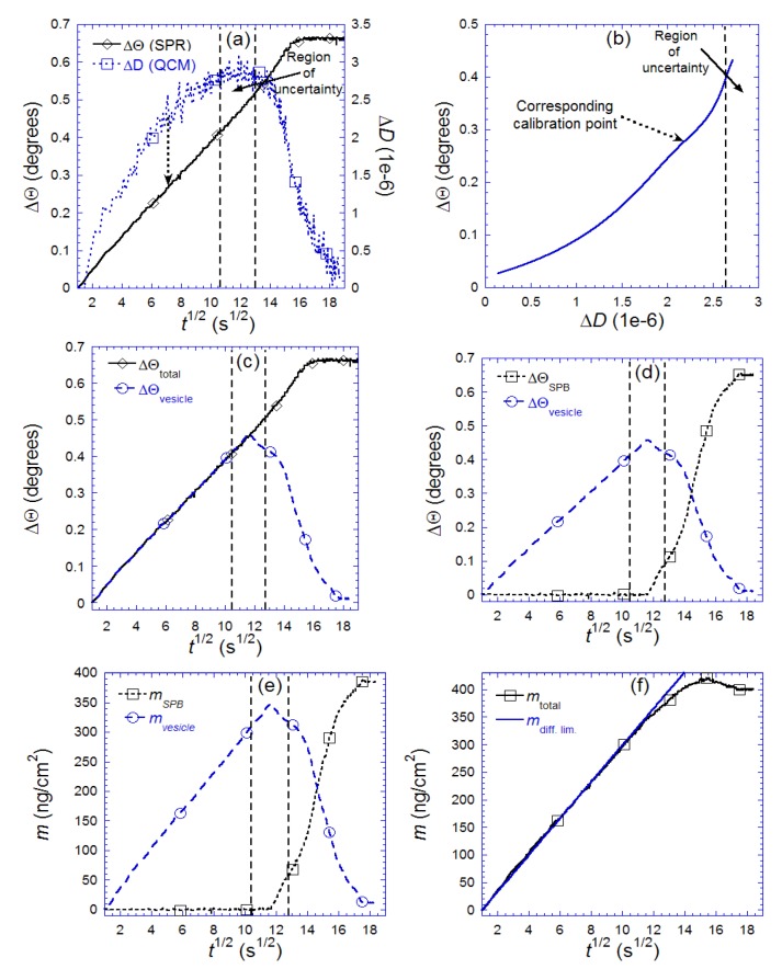
Figure 9.
Illustration of the step-by-step procedure (a–f described in the text) used to separate the contribution to the change in SPR angle, Θ, and coupled mass, mtotal, originating from adsorbed vesicles (ΔΘvesicle, mvesicle) and supported bilayer islands (ΔΘSPB, mSPB), respectively. Also shown in (f) is the expected mass adsorption from diffusion limited transport of lipid material in liposomes. Adapted with permission from Reimhult et al. [14]. Anal. Chem. 2004, 76, 7211–7220. Copyright 2004 American Chemical Society.

