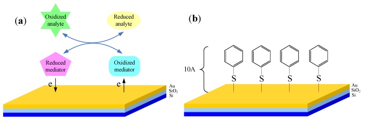
Figure 8.
Schematic diagram of the interface between biological analyte and EMIS structured sensor. (a) Redox couples functioning as mediators used to be added into samples, which usually can be Fe(III)/Fe(II) or quinhydrone; (b) Modified interface using electron transport promoter to accelerate electron transport between analyte and sensor surface, here 4-PySH is taken as an example.

