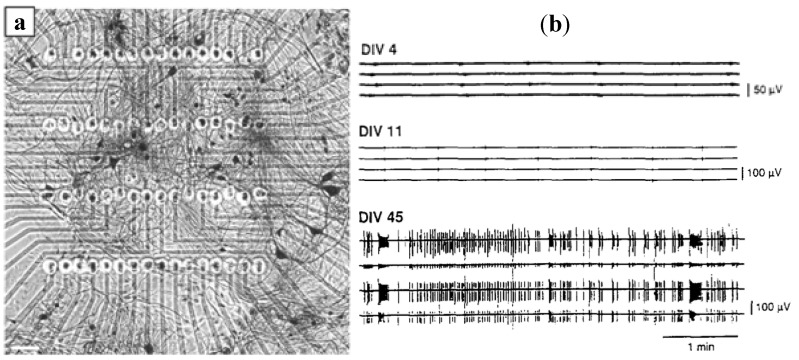
Figure 13.
(a) Neuronal network derived from murine spinal cord tissue (92 days in vitro), grown on the recording matrix of a 64-electrode array plate. (Reprinted from [70]. ©2007, with permission from Elsevier) (b) Developmental changes in neuronal activity. Random bursts were observed at DIV4, tightly synchronized activity appeared at DIV11. The mature neuronal activity consisted of a complicated, high order pattern of spike-like firing and bursting at DIV45. (Reprinted from [71]. © 1996, with permission from Elsevier).

