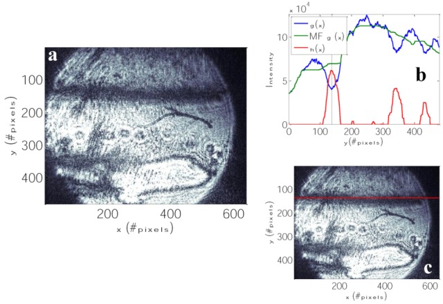
Figure 7.

(a) Reflected images from DBSPRI sensor of 11-MUA + (NHS and DCC) after immobilization of estrone antibody and attached to it the estrone and after washing with PBS; (b) detection steps using the DBSPRI extraction algorithm [32]: blue—g(x), which is integrated original image; green—median filtered integrated image’ and red—median filtered image after background suppression. SPR line location is pointed by vertical lines; (c) reflected light, which was captured by the camera using the optical arrangement described in Figure 3, with extracted locations of the SPR signal, indicated by a solid line along the image.
