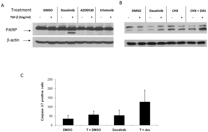
Figure 3. Induction of apoptosis after treatment with TGFβ-1 and dasatinib.
(A) A549 cells were treated with 100 nM of dasatinib, 1000 nM of AZD0530, or erlotinib with or without 5 ng/mL TGFβ-1 for 48 hours. (B) A549 cells were pre-treated with 10 µg/mL cycloheximide (CHX) for 1 hour, followed by TGFβ-1 plus or minus dasatinib. After incubation, cells were harvested, lysed, and PARP cleavage detected by Western Blot analysis. (C) A549 cells were seeded in 96-well plates at 5×10³ per well. Cells were treated, and Cell Player 96-Well Kinetic Caspase 3/7 Reagent was added simultaneously. Treatments were done in triplicate. Values are shown as the average number of caspase 3/7 positive cells from 3 independent experiments.

