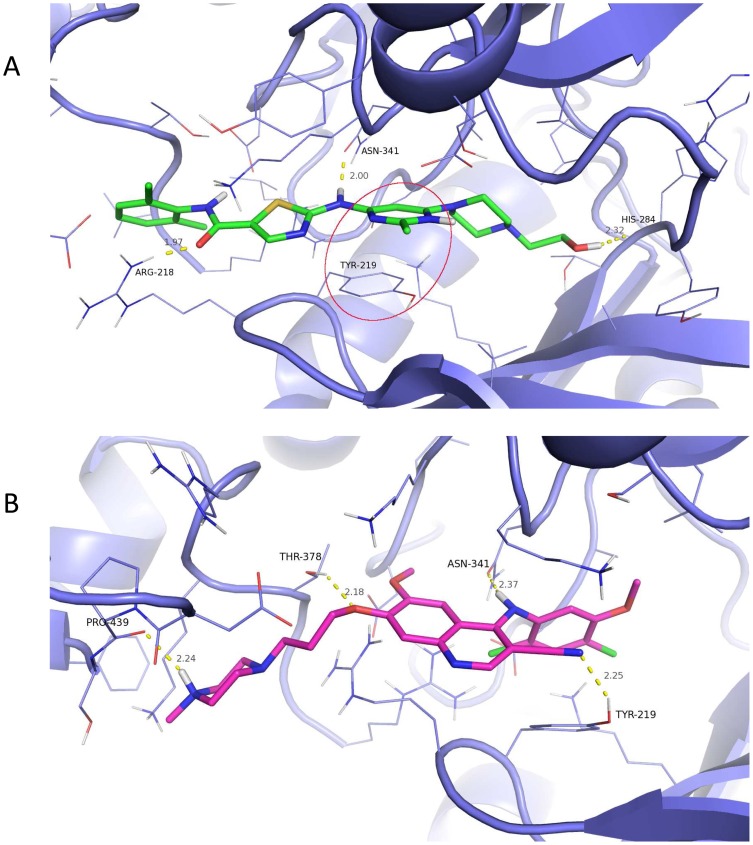
Figure 7. 3D Docked Pose and Interactions of dasatinib and bosutinib with TβR-I 3D rendering of the binding pose of (A) dasatinib and (B) bosutinib docked into TβR-1.
Protein is represented by purple carbon cartoon representation with residues surrounding a ligand are in line representation. Bosutinib is colored with magenta carbons and dasatinib with green carbons. Hydrogen bonds represented by dashed yellow lines with distances (gray) and interacting residue (black) labeled. A π-π stacking interaction between the phenyl of TYR-219 and the pyrimidine of dasatinib is circled in red.

