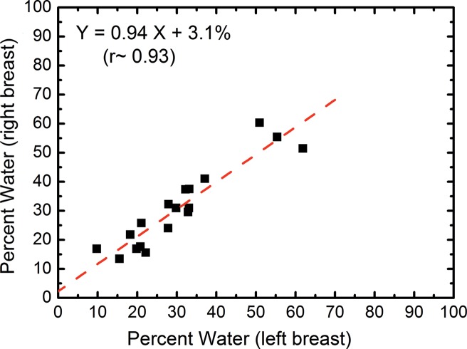
Figure 4a:
Scatterplots show the right-left correlation of (a) water and (b) lipid contents measured from spectral CT by using dual-energy decomposition and (c, d) from chemical analysis. Linear fittings for all three contents are shown with dashed lines, which are in good agreement with the identity line.

