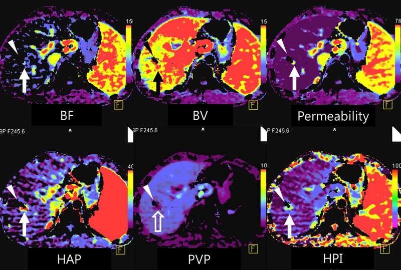
Figure 9b:
Images show perfusion CT facilitating diagnosis of local recurrence after TACE for HCC. (a) On precontrast (top left), arterial (top right), portal venous (bottom left), and delayed (bottom right) phase perfusion CT images 30 days after treatment, a densely lipiodol-laden nodule (arrow) is in right lobe of liver. No definite enhancing viable tumor focus is seen on conventional CT scans. (b) Color maps of perfusion CT, however, show area (arrows) of increased blood flow (BF) (top left), blood volume (BV) (top middle), permeability (top right), HAP (bottom left), and HPI (bottom right) and decreased PVP (bottom middle), indicating recurred/residual HCC at leading edge of tumor (arrowheads). (c) Arterial phase image (left) obtained 1 month after perfusion CT shows increased size of enhancing viable tumor (arrow) around faint lipiodol residuals (arrowhead), with washout on portal venous phase image (right), confirming recurrent/residual disease.

