
Figure 3b:
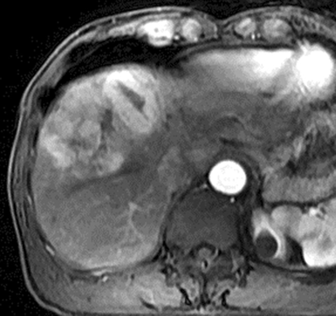
Images in 64-year-old man with HCC and hepatitis B–related cirrhosis: multiphasic MR technique with extracellular contrast agent. (a–d) Gadolinium-enhanced T1-weighted three-dimensional (3D) gradient-echo (GRE) images (repetition time msec/echo time msec, 6.3/1.9; flip angle, 15°) show large hypointense mass on (a) precontrast image with (b) hyperenhancement in late hepatic arterial phase. Notice enhancement of portal vein branches but not of hepatic vein branches in late hepatic arterial phase. (c) Portal venous and (d) 3-minute delayed phase images show persistent enhancement of tumor relative to liver. Persistent enhancement is atypical of large progressed HCCs, which characteristically appear to washout on venous phase images. Notice capsule appearance (arrow) on delayed phase image. Capsule appearance is seen to better advantage on delayed compared with portal venous phase image and permits confident diagnosis of HCC despite lack of washout appearance.