Figure 5c:

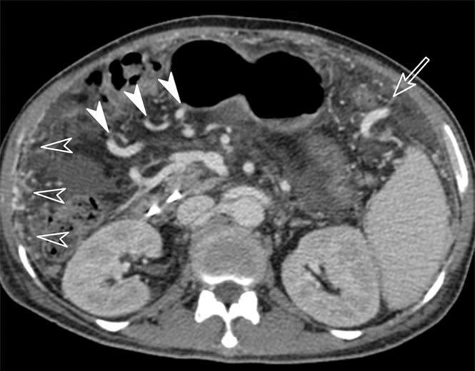
Images obtained in a 61-year-old man with hepatitis B virus–associated liver cirrhosis. (a) MR elastogram shows the mean stiffness of the liver to be 9.3 kPa and mean stiffness of the spleen to be 8.6 kPa. (b) Endoscopic image demonstrates a grade 1 esophageal varix. (c, d) Axial CT scans obtained in the portal venous phase demonstrate that the patient has multiple collateral veins, such as omental (open arrowheads on c), perisplenic (open arrow in c), mesenteric (solid arrowheads on c and d), and retroperitoneal (solid arrow in d) varices.
