
Figure 1c:
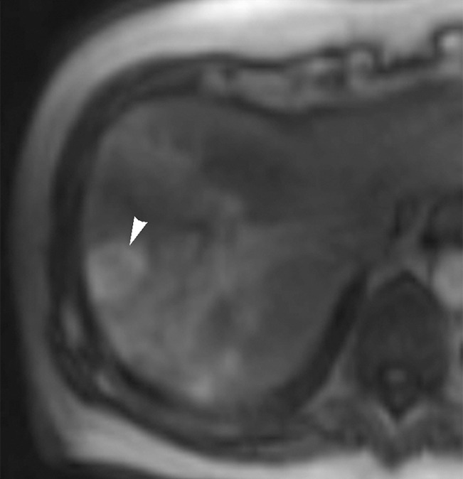
Images in 65-year-old man with focal HCC (arrowhead). (a) Postcontrast arterial phase T1-weighted MR image shows 2.2-cm tumor. (b) Segmental chemoembolization of hypervascular mass was subsequently performed. (c) Pre- and (d) postchemoembolization TRIP MR images demonstrate marked tumor perfusion reduction, which was calculated to be 69%. Clinical follow-up revealed 22.7-month TFS.