
Figure 2b:
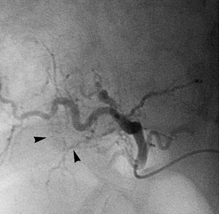
Images in 54-year-old man with nodular HCC (arrowheads). (a) Postcontrast arterial phase T1-weighted MR image depicts 2.4-cm enhancing mass. (b) Chemoembolization of tumor was consequently performed. (c) Pre- and (d) postchemoembolization TRIP MR images display limited tumor perfusion reduction, which was calculated to be 31%. Clinical follow-up demonstrated 5.9-month TFS.