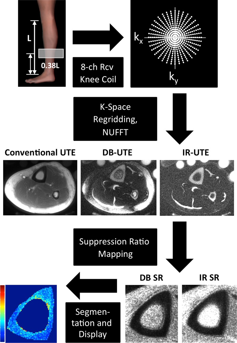
Figure 3:
Flowchart of SR data acquisition and processing procedure shows an axial section acquired without (Conventional UTE) and with dual-band saturation pulse and adiabatic inversion pulse suppression. SR maps were calculated by obtaining the ratios between a conventional UTE image and the corresponding DB-UTE and IR-UTE images. After segmentation of the periosteal and endosteal cortical boundaries, SR parametric maps of the cortical bone can be obtained. 8-ch Rcv = eight-channel receive, NUFFT = nonuniform fast Fourier transform.

