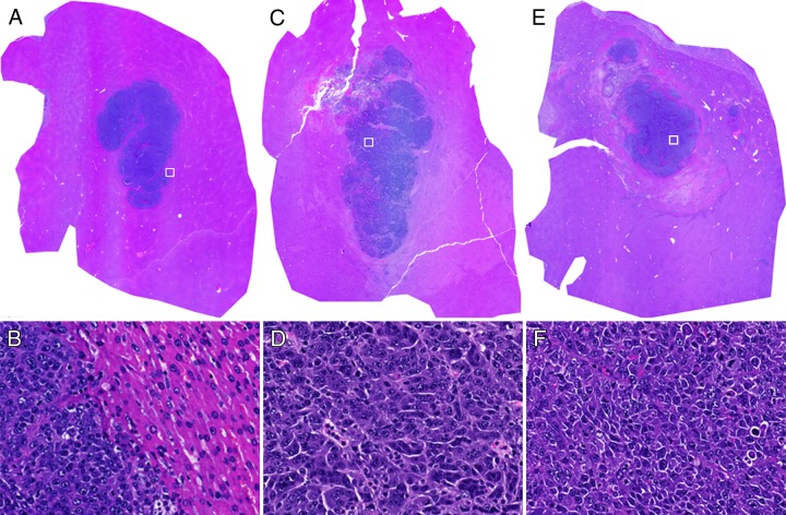
Figure 3:
Representative hematoxylin-eosin–stained histologic slides from tumor tissues in, A, B, control rats, C, D, rats sacrificed 30 minutes after IRE, and E, F, rats sacrificed 24 hours after IRE. Nucleus agglutinations can be observed 30 minutes after IRE. Tumor cells are arranged more loosely in F than in B. The nucleus-to-cytoplasm ratio tended to increase 24 hours after IRE. □ = approximate locations for D, E, and F. Original magnifications are ×10 for A, B, and C and ×40 for D, E, and F.

