
Figure 2:
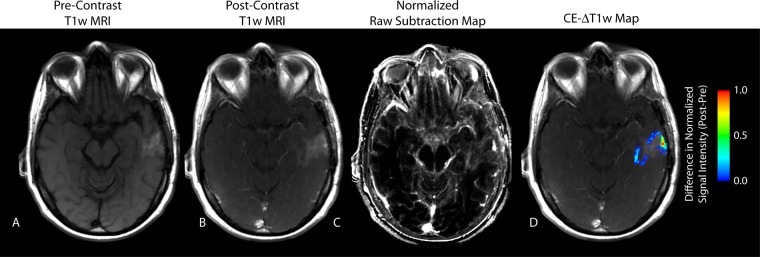
Nonenhanced T1-weighted MR image in a 50-year-old male patient with recurrent GBM demonstrates significant T1 shortening after bevacizumab therapy, which may have been mistaken as residual tumoron CE T1-weighted MR image. A, Nonenhanced T1-weighted MR image. B, CE T1-weighted MR image. C, Subtraction map. D, CE T1-weighted subtraction map shows demarcation of an area of residual tumor identified as a positive increase in MR signal intensity after administration of contrast agent. Keys are the same as on Figure 1.