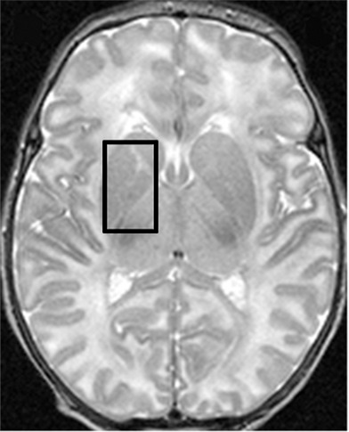
Figure 4a:

1H MR spectroscopy in early assessment of perinatal hypoxia-ischemia in the newborn. MR imaging was performed (a–c) 12 hours and (d, e) 10 days after perinatal asphyxia. (a) Axial T2-weighted image shows no signal abnormalities, whereas (b) spectrum (1.5 T, PRESS, 1500/288, 192 repetitions) obtained from the right basal ganglia shows markedly increased Lac resonance with preserved tNAA, tCr, and tCho resonances. (c) Diffusion-weighted image (echo-planar imaging; 30 directions; b value, 700 sec/mm2) with axial apparent diffusion coefficient map shows no diffusion abnormalities. (d) Axial T2-weighted images show areas of high (white arrows) and low (black arrows) signal intensity in putamen and thalamus, representing clear ischemohemorrhagic lesions. (e) Axial proton density images demonstrate prominent detection of lesion’s extension. (Reprinted, with permission, from reference 76.)
