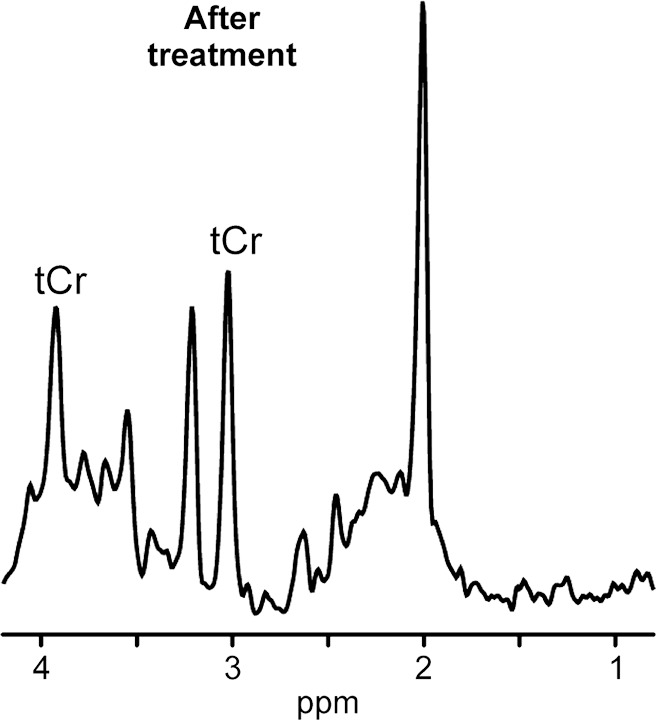
Figure 5b:

1H MR spectroscopy of neurometabolic disorder. (a, b) White matter spectra (1.5 T, PRESS MR spectroscopic imaging, 3000/30, six weighted averages, nominal voxel size = 10 × 10 × 15 mm3) in girl with guanidinoacetate methyltransferase deficiency before treatment at age 3 years 2 months (a) and after 3.5 months of treatment with oral creatine supplementation (b). Resonance from creatine-containing metabolites (tCr) returned to normal in this region as well as in other investigated brain areas.
