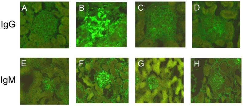
Figure 2. Immunofluorescence microscopy.
Glomerulus from a normal control mouse (A, E), model control mouse (B, F), resveratrol A mouse (C, G), and resveratrol B mouse (D, H). Panels A, B, C, and D were stained with goat polyclonal anti-mouse IgG-FITC and panels E, F, G, and H were stained with goat polyclonal anti-mouse IgM-FITC.

