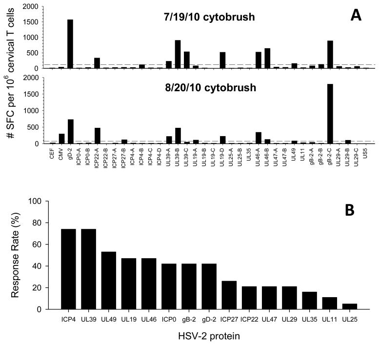
Figure 2. Antigenic diversity of HSV-2 specific T cells in cervical T cell lines.
(A) Cervical T cell lines that were positive by HSV-LP from HSV-2+ Participant 17 obtained on 7/19/10 (top) and 8/20/10 (bottom) were stimulated with HSV-2, CEF or CMV peptide pools and tested for reactivity by IFN-γ ELISPOT; Y-axis is net spot forming cells (SFC) per million cervical T cells (SFC in DMSO control wells were subtracted). Dashed lines represent the threshold for positivity (4X median of DMSO control wells and ≥55 SFCs per million cervical T cells). (B) Cervical T cell lines from HSV-2+ participants that were positive by HSV-LP were screened for reactivity using the 34 peptide pools representing 16 HSV-2 proteins by IFN-γ ELISPOT. The frequency of detecting HSV-2 protein specific T cell responses in one or both cervical cytobrush samples from 19 HSV-2+ participants is displayed: HSV-2 proteins are stratified based on their hierarchy in % response rate.

