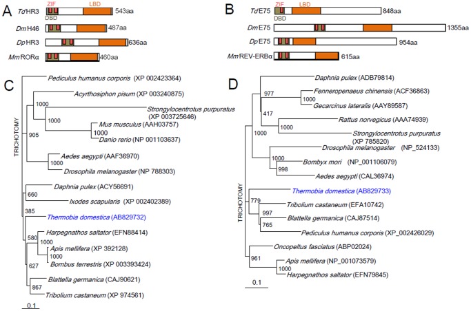
Figure 1. Sequence alignments of conserved domains and phylogenetic neighbor-joining tree of HR3 and E75.
(A, B) Schematic structure of various HR3 (A) or E75 (B) proteins, comparing the organization of the 3 conserved domains, DNA-binding domain (DBD), zinc finger domain (ZIF), and ligand binding domain (LBD). The numbers on the right indicate the number of amino acid residues. Td, Thermobia domestica; Dm, Drosophila melanogaster; Dp, Danaus plexippus; Mm, Mus musculus. (C, D) A phylogenetic neighbor-joining tree of known animal HR3 or E75 proteins. The GenBank or RefSeq accession numbers are indicated in brackets. A reference bar indicates distance as the number of amino acid substitutions per site.

