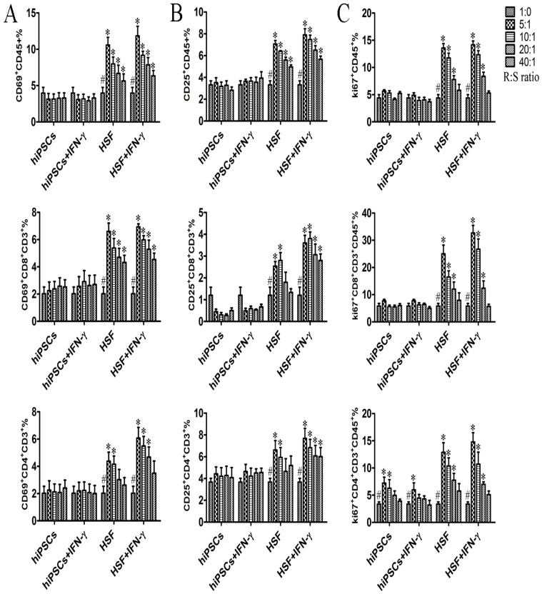
Figure 4. hiPSCs do not effectively induce activation and proliferation responses on allogeneic lymphocytes.
hiPSCs pretreated with or without IFN-γ (stimulator cells; S) were inactivated and then directly cultured with allogeneic PBMCs (responder cells; R) at different R/S cell ratios in a MLR for 5–7 days (n = 4). PBMCs were harvested for examination of the expression of activation markers and Ki67 protein at the indicated time point. Surface expression of CD69 and CD25 on PBMCs, CD3+CD8+ T cells, and CD3+CD4+ T cells was measured by flow cytometry after 6 h and 24 h of co-culture (A, B). (C) Intranuclear Ki67 protein expression in PBMCs, CD3+CD8+ T cells and CD3+CD4+ T cells was analyzed after 5–7 d of stimulation. Data are shown as the mean ± SEM. Results are representative of four different experiments. #, indicating significant difference compared to those with different ratio of stimulator within same group (p<0.05). *, indicating significant difference compared to those with same and same ratio of stimulator (p<0.05).

