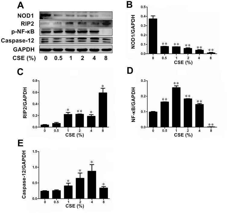
Figure 1. CSE exposure altered levels of NOD1, Caspase12, RIP2, and p-NF-κB protein expression in Leuk-1 cells.
A Immunoblot bands indicated effects of CSE exposure on protein expression of NOD1, RIP2, p-NF-κB and Caspase12. B CSE treatment decreased the expression of NOD1 in a dose-dependent manner. C CSE treatment increased the expression of RIP2 in a dose-dependent manner. D p-NF-κB expression was activated by 0.5%∼4% CSE exposure, while 8% CSE treatment reduced p-NF-κB levels significantly. The p-NF-κB expression reached the highest level in Leuk-1 cells following 1% CSE exposure. E Caspase12 expression was activated by 1%∼8% CSE. Immunoblot band density data were expressed as means±SE (n = 3). Statistical significance: *P<0.05, **P<0.01 vs. cells without CSE treatment.

