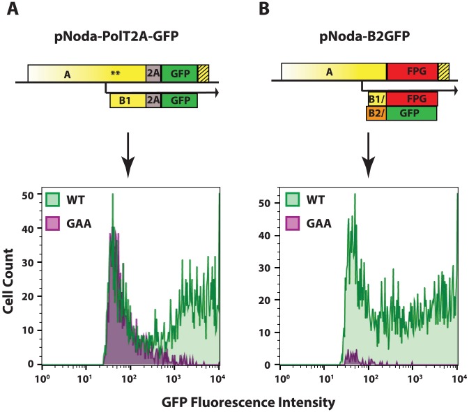
Figure 5. GFP expression from the two replicon designs.
A. Protein A fusion replicon construct and FACS analysis, B. Protein B2 fusion replicon construct and FACS analysis. The hatched yellow box represents the extension of the RNA element, as in Fig. 3. 48 hours after transfection into BSR, cells were trypsinized and examined by FACS. Cells in the GFP-positive gate are shown. Cells transfected with wt polymerase constructs are shown in green; those transfected with GAA mutant polymerase constructs are shown in purple.

