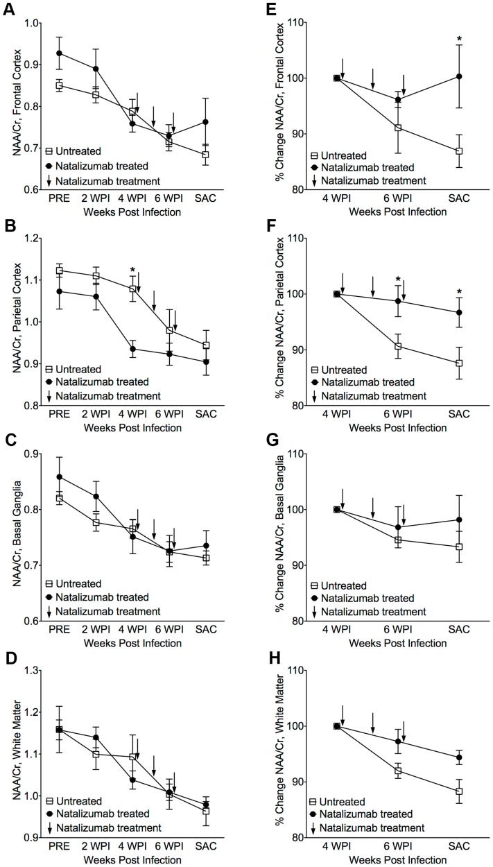
Figure 2. Stabilization of neuronal injury in SIV-infected animals following natalizumab treatment.
(A) A decreased NAA/Cr ratio in frontal cortex (FC), (B) parietal cortex (PC), (C) basal ganglia (BG), and (D) white matter (WM), was observed in untreated and natalizumab treated animals by four weeks post infection (WPI). Decreased NAA/Cr stabilized with natalizumab treatment (indicated by arrows at 28, 35, and 42 dpi) in the FC (E), PC (F), BG (G), and WM (H). Each point represents the mean ± SEM. P≤0.05* using Holm-Šídák post-tests following significant repeated measures ANOVA.

