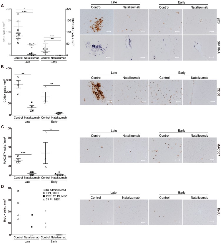
Figure 3. Natalizumab treatment blocks the traffic and accumulation of SIV infected monocyte/macrophages in the brain.
(A) Natalizumab treatment on 28 dpi (late), resulted in scattered SIV p28+ and RNA+ cells in the CNS (frontal cortex, parietal cortex, occipital cortex, brainstem) relative to controls, all sacrificed with progression to AIDS. Natalizumab at the time of infection (early) prevented the traffic of SIV p28+ and RNA+ cells into the parenchyma, while several SIV p28+ and RNA+ cells were evident in tissues of untreated controls that were all sacrificed on 22 dpi. (B) Late natalizumab treatment resulted in decreased numbers of CD68+ macrophages in the brain relative to controls. Numbers of CD68+ macrophages were significantly reduced in the brains of early natalizumab treated animals compared to matched controls. (C) Fewer MAC387+ cells were observed in late natalizumab treated macaques compared to non-treated animals. Significantly less MAC387+ monocytes were detected in early treated macaques than in untreated controls. (D) To determine the timing of monocyte/macrophage egress into the CNS, animals were administered BrdU at various time points. In animals given BrdU after late natalizumab treatment had begun (33 dpi, 24-hours prior to necropsy), there were no BrdU+ monocyte/macrophages any brain region examined. Lower numbers of BrdU+ cells were detected in natalizumab treated than non-treated animals that all received BrdU throughout infection (−9 dpi, 26 dpi, and 24-hours prior to necropsy). No recently recruited BrdU+ monocyte/macrophages were found in the parenchyma of animals treated with early natalizumab. Scale bars: 50 microns. P values calculated using unpaired t tests. P≤0.05*, p≤0.01**, p≤0.001 ***.

