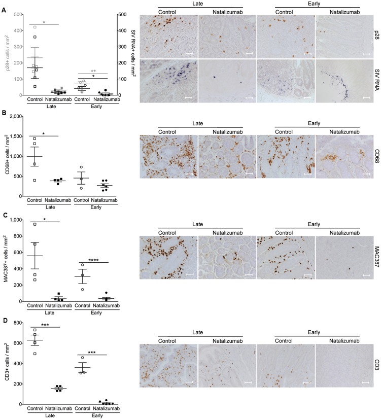
Figure 4. Natalizumab reduces accumulation of productively infected cells, CD3+ T cells, and MAC387+ monocytes to gut.
(A) There are significantly fewer SIV p28+ infected cells in the duodenum, jejunum, and colon tissues from late natalizumab treated animals compared to untreated animals. The numbers of SIV RNA+ cells were similar between both groups. Less SIV p28+ and RNA+ cells were detected in guts of early natalizumab treated macaques compared to controls. (B) Untreated animals had significantly higher numbers of CD68+ macrophages than late natalizumab treated animals. There were comparable numbers of CD68+ macrophages in gut tissues from early natalizumab treated and non-treated animals. (C) Untreated controls had significantly higher numbers of MAC387+ cells in the intestine than macaques starting natalizumab late in infection. Natalizumab treatment early reduced the number of infiltrating MAC387+ monocytes compared to controls. (D) There were fewer CD3+ T lymphocytes in the gut of late and early natalizumab treated animals compared to controls. Lines and error bars indicate the mean ± SEM for each treatment group. Scale bars: 50 microns. P values calculated using unpaired t tests. P≤0.05*, p≤0.01**, p≤0.001 ***, p≤0.0001 ****.

