
Fig. 11.
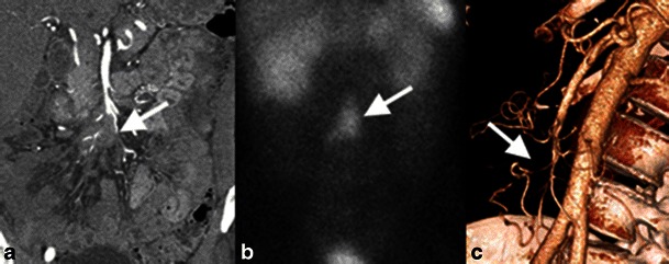
Mesenteric ischaemia secondary to vascular compression by a small bowel carcinoid. a Coronal contrast-enhanced reformatted image shows mesenteric metastatic disease from the small bowel carcinoid encasing distal branches of the SMA (white arrow). b Coronal image from an octreotide scan shows uptake by the metastatic disease (white arrow), indicating tumoral somatostatin receptors. c Three-dimensional volume-rendered reconstruction shows attenuated calibre of the ileocolic artery (white arrow)