
Fig. 12.
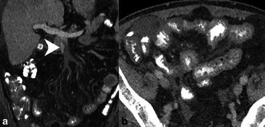
SMV thrombosis. a Contrast-enhanced coronal reformatted CT demonstrates occlusive thrombus in the SMV near the portal confluence (white arrowhead). b Axial CT image demonstrates diffuse small bowel wall thickening secondary to venous congestion and ischaemia