
Fig. 13.
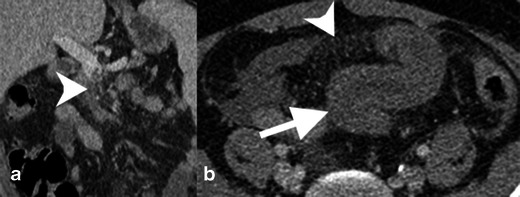
SMV thrombosis with mesenteric congestion. a Contrast-enhanced coronal reformatted CT of an occlusive thrombus in the SMV (white arrowhead) extending to the portal confluence. b Axial contrast-enhanced CT image demonstrates mesenteric congestion with engorged mesenteric veins and trace fluid (white arrowhead). The small bowel is thickened because of the venous stasis and probable ischaemia