
Fig. 14.
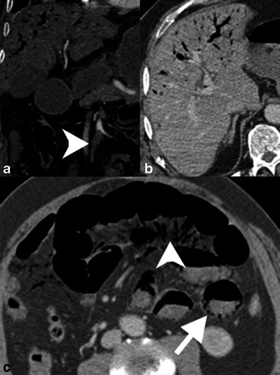
Portomesenteric venous gas. a Contrast-enhanced coronal reformatted CT demonstrates occlusion of the SMA (arrowhead). Portal venous gas is seen in the liver. b Axial CT shows portal venous gas in the anti-dependent liver. c The small bowel demonstrates luminal dilation, a paper-thin wall and poorly enhancing mucosa. There is mesenteric venous gas (arrowhead) as well as pneumatosis in the dependent bowel wall. The patient had established bowel necrosis and died shortly after imaging