
Fig. 5.
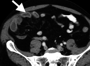
Hyper-enhancement of the bowel mucosa. Axial contrast-enhanced CT image demonstrates hyper-enhancement of the right lower quadrant ileal mucosa (arrow) secondary to superior mesenteric artery occlusion (not shown). The degree of mucosal enhancement can be compared to left-sided bowel loops as an internal control