
Fig. 8.
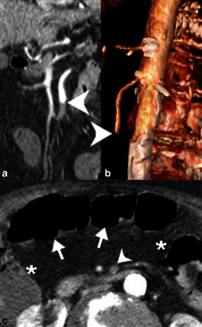
Embolic occlusion of the superior mesenteric artery. a Contrast-enhanced coronal reformatted CT image and b 3D volume-rendered reconstruction of demonstrate an abrupt cutoff of the proximal superior mesenteric artery (white arrowheads) secondary to an embolus. c Axial contrast-enhanced CT demonstrates relatively poor enhancement of the transverse colon (white arrows) relative to the hepatic and splenic flexures (*). Note also the occluded SMA (white arrowhead)