
Fig. 9.
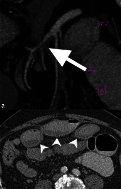
SMA thrombosis. a Contrast-enhanced, coronal reformatted CT images demonstrate an eccentric filling defect in the proximal SMA, corresponding to thrombus formation. b Axial contrast-enhanced CT image shows oedematous bowel wall thickening of small bowel loops (white arrowheads) and mesenteric congestion. This patient subsequently improved with conservative anticoagulation therapy