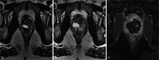
Fig. 19.

Contralateral treatment. A 56-year-old patient, with initial right photodynamic therapy, then left therapy in a second intervention. Pre-left therapy (a) and day-7 post-left therapy (b) axial T2-weighted images; day-7 post-left therapy (c) axial T1 FS contrast-enhanced MR image. On the left lobe, a heterogeneous T2 area with low signal in T1-weighted image, unenhanced, corresponding to the recent tissue necrosis (star). In the right lobe the T2 hyperintense old cystic cavity increased in size (arrowhead)
