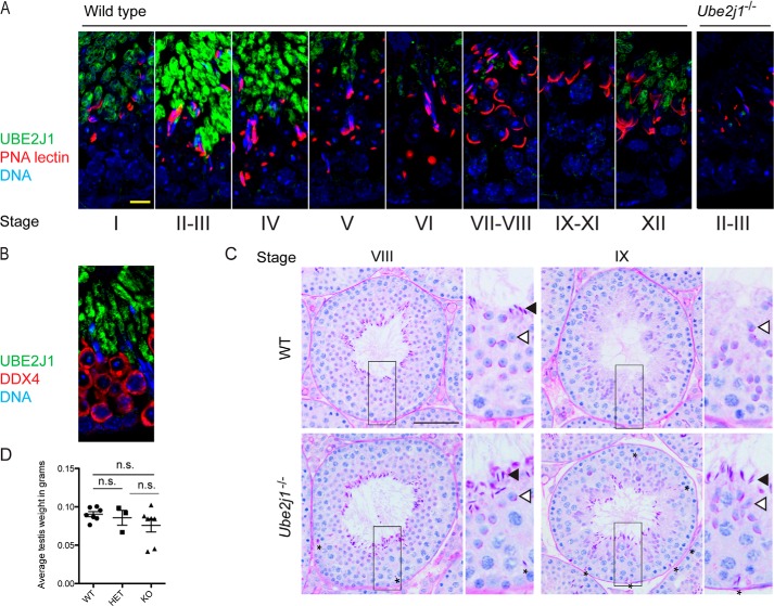
FIGURE 6.
Male Ube2j1−/− mice have defects in spermatid differentiation. A, immunohistochemical staining for UBE2J1 (green), acrosomes (PNA lectin, red) and DNA (DAPI, blue) in testis cross-sections of a wild type mouse. Note the stage-specific expression of UBE2J1. No staining was detected in Ube2j1−/− testes (right panel). Scale bar, 10 μm. B, immunohistochemical staining for UBE2J1 (green) and the germ cell-specific DDX4 (red) in a wild type testis. C, PAS-stained cross-section of testes from Ube2j1+/+ (WT, top row) and Ube2j1−/− (bottom row) mice. Note the abnormal germ cell associations and spermiation defect at stage IX. Asterisks denote mislocated spermatids. Filled arrowheads, elongated spermatids to be released at the end of stage VIII. Empty arrowheads, next generation round spermatids. D, average testis weight in grams of Ube2j1+/+ (WT), Ube2j1+/− (HET), and Ube2j1−/− (KO) mice. Shown are mean testis weight of individual mice (data points), and overall average with standard deviation. Not significant (n.s.) indicates p > 0.05, as determined by one-way ANOVA with Tukey's Multiple Comparison Test.

