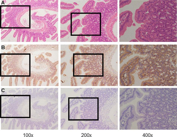
FIGURE 2.
Immunohistochemistry on sections obtained from mouse duodenal mucosal tissues. A, H&E staining of mouse duodenal mucosa showing normal morphology and typical villous crypt structures. B, representative immunoreactivity of CaSR proteins (brown) in the villus and crypts of the duodenal mucosa at different magnifications. C, representative immunoreactivity without incubation of primary anti-CaSR antibody in the villus and crypts of duodenal mucosa as a negative control. Magnifications are ×100, ×200, and ×400 in the left, center, and right panels, respectively. These data are representative of at least three experiments with similar results.

