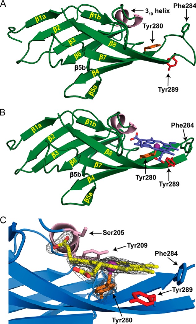
FIGURE 6.

Crystal structures of apo- and holo-Hbp2N2. A and B, ribbon representation of apo-Hbp2N2 (A) and holo-Hbp2N2 (B). The β strands and helices are colored green and light pink, respectively. Residues are color-coded to match the sequence alignment in Fig. 1C. Secondary structural elements are labeled as described in the text. The hemin molecule is colored blue. C, the hemin binding pocket is shown with the 1.8-Å resolution holo-Hbp2N2 structure superimposed with a simulated annealing Fo − Fc omit map contoured to 5.0 σ shown as a dark gray mesh.
