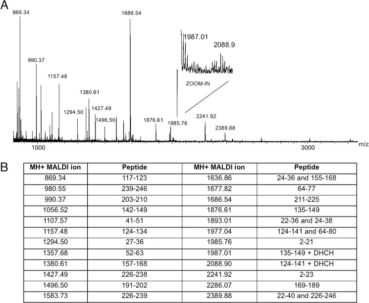
FIGURE 10.
Identification of reactive arginines in phosphomannomutase2 monitored by MALDI mass spectrometry. A shows the MALDI mass spectrum of WT-PMM2 incubated with 1,2-cyclohexanedione (molar excess of 100) for 140 min and digested by trypsin for 90 min. B shows the list of MH+ values measured by MALDI MS and attributed to tryptic peptides. Values at 1987.01 and 2088.90 atomic mass units were compatible with peptides 135–149 and 124–141 alternatively modified by 1,2-cyclohexanedione at Arg141 and Arg134, respectively.

