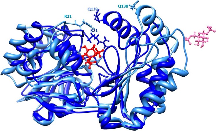
FIGURE 2.
Protein closure along ligand binding. The initial model in open conformation is shown in pale blue, and the model in closed conformation is shown in dark blue. Side chains of two residues that come close upon domain closure, Arg21 and Gln138, are shown as sticks. The initial and final positions of the ligand are shown in pink and red, respectively, with a ball and stick representation. The images were drawn with Chimera (27).

