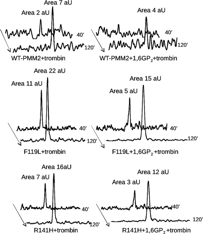
FIGURE 7.

Limited proteolysis of wild type, F119L, or R141H phosphomannomutase2 with thrombin monitored by mass spectrometry. The extracted ion chromatograms relative to the formation of peptide 2–21 are reported for wild type (A and B), F119L (C and D), and R141H (E and F) phosphomannomutase2. Samples were incubated in the absence (A, C, and E) or presence (B, D, and F) of α-Glc-1,6-P2 (1,6GP2) with thrombin for 40 or 120 min. Peak areas were calculated by MassLynx 4.0 software and are reported as arbitrary units (aU).
