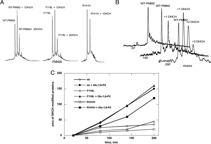
FIGURE 9.
Chemical modification of reactive arginines in phosphomannomutase2 monitored by mass spectrometry. A shows deconvoluted RP-LC-MS mass spectra of WT-PMM2, F119L, and R141H incubated with 1,2-cyclohexanedione (molar excess of 100) for 140 min, revealing the different modification levels of the three proteins. B shows the time course modification of WT-PMM2, revealing unmodified protein (molecular mass of 27,950.25 Da) and mono- and di-modified species (molecular masses of 28,061.50 and 28,173.10 Da). C shows the graph of the area of the peaks of deconvoluted spectra relative to 1,2-cyclohexanedione-modified WT-PMM2, F119L, and R141H with or without Glc-1,6-P2 measured at different incubation times. ′, minutes.

