
FIGURE 7.
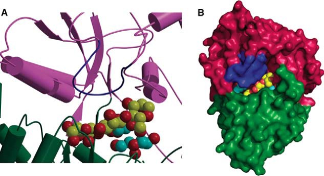
Model for binding of branched substrates by CpMnBP1. Shown is a close-up view of the active site (A) and the solvent-accessible surface (B) of a hypothetical model of a branched mannotriose ligand bound in the ligand-binding pocket of CpMnBP1. The oligosaccharide is shown in Corey-Pauling-Koltun representation with the branching carbon atoms colored in cyan.