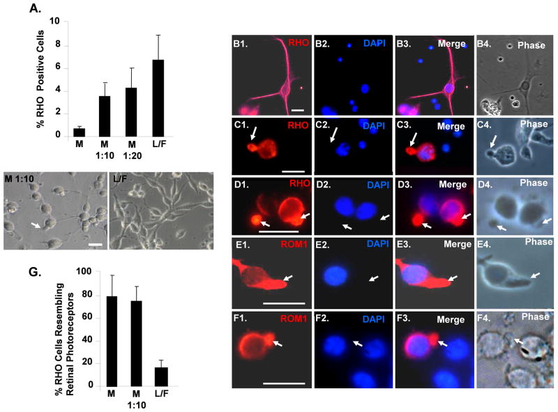
Fig 4.
Adherent culture on Matrigel enhances the number of differentiating iPSC resembling primary cultures of rod photoreceptors. (A): Quantification of RHO+ cells generated on different matrices. “L/F” laminin/fibronection, “M” Matrigel. Phase images of cells are shown below. P<0.05 for all values compare to undiluted Matrigel. (B1–B4): Differentiated iPSC plated on laminin/fibronection and immunostained for RHO. Note cells with elongated cell bodies that immunostain uniformly for RHO. (C1–C4): Differentiated iPSC cells plated on Matrigel and immunostained for RHO. Note that RHO is concentrated in projections from the main cell body. (D1–D4): Primary culture of swine retinal cells immunostained for RHO. Note that RHO is concentrated in outer segments extending from the rounded cell bodies, and that the cell morphology resembles that of the differentiated iPSC plated on Matrigel in panels C1–C4. (E1–E4): Differentiated iPSC plated on Matrigel and immunostained with the rod outer segment marker ROM1. Note that ROM1 colocalizes with RHO in projections from the rounded cell bodies. (F1–F4): Primary culture of swine retinal cells immunostained for ROM1. Note that, like RHO, ROM1 is concentrated in outer segments projected from the rounded cell bodies and that these cells resemble those in panels E1–E4. Scale bars = 20 μm. (G): Quantification of RHO+ iPSC morphologically resembling primary culture rod photoreceptors generated on different matrices. P<0.05 for L/F compared to M and M 1:10.

