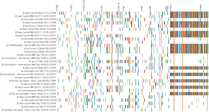
Figure 1. Distribution of polymorphic residues in the proteome of North American avian H1N1 IAVs.
The proteome was concatenated using the polymorphic sites in each protein in the following order: PB2, PB1, PB1-F2, PA, PA-X, HA, NP, NA, M1, M2, NS1, and NEP. Similar amino acids were grouped by color: aliphatic amino acids, shades of brown and orange; positively charged residues, shades of blue; negatively charged ones, shades of red; polar uncharged ones, shades of green; aromatic ones, shades of purple; and glycine and cysteine, shades of yellow. These positions were based on the sequences used in this study.

