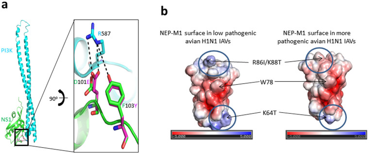
Figure 5. NS1 and NEP variants associated with pathogenicity by residue effect.
(a) Ribbon model of NS1 (green) in which S103 is replaced with F103, and NS1 is bound to the PI3K inhibitory domain (teal) [Bovine PDB: 3L4Q] are labeled. The predicted interaction (black dots <5 Å) between variant residues E101 and Y103 of NS1 protein with R587 (numbering is based on human) from the PI3K inhibitory domain. Low-pathogenic strains contain residue D101 and F103 in which D101 interacts only with the PI3K inhibitory domain R587. Pathogenic viruses contain E101 and Y103, and both residues are involved in interactions. The outcome is a stronger interaction between NS1 and the PI3K inhibitory domain. (b) NEP variants increase the negative charge of the M1-binding domain in more pathogenic viruses. Electrostatic surface potential representation of the NEP/M1-binding surface [PDB: 1PD3]; native (left image) and with three variant changes (R86I, K88T, and K64T) (right image). The surface color indicates negative charge (red), positive charge (blue), and neutral (white). The extent of the charge is indicated below each image in units of kT/e. These positions were based on the sequences used in this study.

