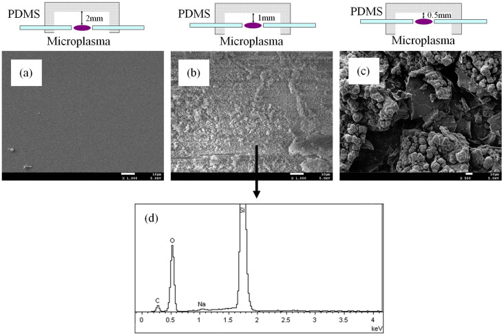
Figure 3. Influence on the PDMS substrate.
SEM characterization on the inner walls of the chambers with a distance of (a) 2 mm, (b) 1 mm, and (c) 0.5 mm to the microplasma discharge centers after working of 2 h. The scale bar in the SEM picture is 10 μm. (d) Elemental characterization for the deposition on the inner wall of the chamber (b) by EDX spectrum.

