
Figure 6.
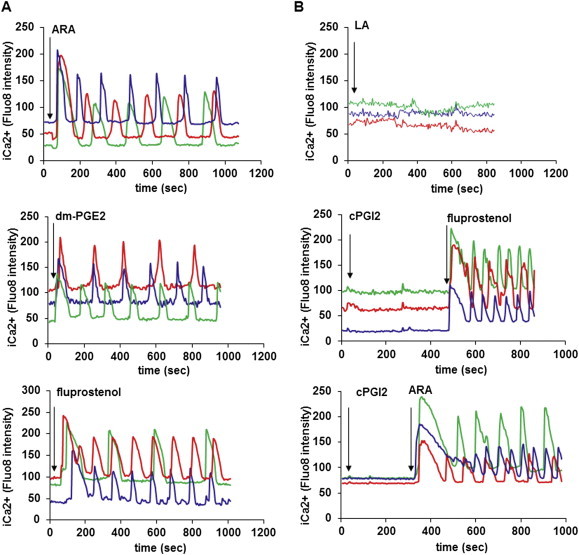
Intra-cellular Ca++ oscillations are induced by ARA and its metabolites. hMADS cells were differentiated into brite adipocytes in the presence rosiglitazone and incubated 15 min with a fluorescent sensitive Ca++ probe (Quest Fluo-8). Cells were analyzed by live fluoromicroscopy. (A) 100 μM ARA, 10 μM dm-PGE2 and 100 nM fluprostenol induce a transitory increase of i[Ca++], followed by i[Ca++] oscillations with a sustained frequency and intensity. (B) 100 μM LA and 10 μM cPGI2 did not trigger i[Ca++] rise. Each track represents integrated imaging of an individual cell. These data are representative of 5 independent experiments (10–50 cells recorded in each experiment).