
Figure 1.
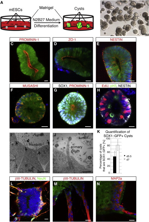
Characterization of Neural Cysts Derived from mESCs
(A–G) Scheme of the generation of neural cysts. Within 5 days neuroepithelial cysts form that express SOX1::GFP and possess a single lumen (B). Day 6 neural cysts are apicobasally polarized, as shown by the luminally expression of PROMININ-1 (C and G) and the tight junction marker ZO-1 (D). The neural stem cell markers NESTIN (E) and MUSASHI (F) are also being expressed. To show that SOX1::GFP cysts are 100% neural in character, we also stained for the protein SOX1 (white nuclei, G).
(H) Cells in a cyst undergo interkinetic nuclear migration, as evidenced by staining for phosphorylated histone 3 (pH3) and the thymidine analog EdU. Cells undergoing division are located at the apical side (pH3+), whereas cells in S phase which are EdU+ are located at the basal side.
(I and J) Electron microscopic analysis of early neuroepithelial cysts. Cysts possess tight junctions (arrows), have large apical membrane surfaces with microvilli and primary cilia (J), and shed midbodies (stars) into the lumen.
(K) Quantification of SOX1::GFP+ cysts at day 5.5 and day 7 of differentiation. Data are represented as mean ± SD (n = 3 independent experiments with 100 cysts counted per experiment).
(L–N) Around day 7 to day 8, postmitotic neurons start to grow out basally, as evidenced by NeuN (L) and βIII-TUBULIN (L and M) as well as MAP2a (N).
Nuclei were counterstained with Hoechst. All data shown were derived with the SOX1::GFP reporter cell line 46C. All immunofluorescence panels (C–H and L–N) represent confocal images of 3D cysts. Scale bars represent 20 μm (C–H and L–N), 2 μm (I), and 0.5 μm (J). See also Figure S1.